Can a CT Scan Detect Ovarian Cancer?
May 13, 2025
Can a CT scan detect ovarian cancer? Yes, but with important limitations.
CT scans are valuable tools in the ovarian cancer diagnostic process, but they represent just one piece of a complex diagnostic puzzle. Understanding how these scans work — and what they can and cannot show — is crucial for anyone concerned about ovarian cancer.
What Is a CT Scan and How Does It Work?
A CT scan (Computed Tomography) creates detailed cross-sectional images of your body using X-rays. Unlike standard X-rays that capture a single image, CT scanners rotate around you, taking multiple pictures from different angles. A computer then combines these images to create detailed views of your internal organs and tissues.
Sign up to receive vital updates through email, and learn how you can get involved. "*" indicates required fieldsStay informed
For ovarian cancer detection, CT scans can:
- Reveal abnormal masses in the ovaries and surrounding areas
- Show if cancer has spread to other parts of the body
- Help determine the cancer’s stage
- Guide treatment planning and surgical decisions
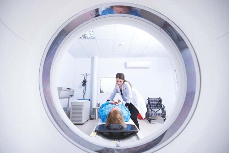
During the procedure, you’ll lie on a table that slides into a donut-shaped machine. The actual scanning typically takes just minutes, though your entire appointment might last longer.
Can You Detect Ovarian Cancer with a CT Scan?
Yes, CT scans can detect ovarian cancer, but they work best when tumors have grown large enough to create visible abnormalities. This represents one of the key challenges in the early detection of ovarian cancer.
Early-stage ovarian tumors are often small and may not appear clearly on CT images. Based on the research materials provided, CT scans have varying sensitivity for detecting peritoneal metastases (cancer spread within the abdomen), with pooled sensitivity rates around 68%.
Does a CT scan detect ovarian cancer in all cases? Unfortunately not. These limitations mean that while CT scans are essential diagnostic tools, doctors typically use them alongside other tests rather than as standalone detection methods.
It’s crucial to understand that while imaging tests like CT scans provide valuable information, they cannot definitively diagnose ovarian cancer. Only a biopsy — removing tissue and examining it under a microscope — can confirm cancer. This is why the diagnostic process often includes multiple steps beyond imaging.
The Role of CT Scans in Ovarian Cancer Diagnosis
Doctors usually order a CT scan after you’ve already shown ovarian cancer symptoms or had abnormal findings on other tests.
CT scans typically fit into the diagnostic process like this:
- You report symptoms to your doctor (persistent bloating, pelvic pain, feeling full quickly, frequent urination)
- Your doctor performs a physical and pelvic exam
- They order an ultrasound (often the first imaging test)
- If these initial tests raise concerns, a CT scan may follow
- Additional tests might include blood tests for tumor markers like CA-125
CT scans excel at “staging” — determining if and how far cancer has spread. This critical information directly shapes treatment planning.
Limitations of CT Scans for Ovarian Cancer
Ovarian cancer CT scans have several important limitations:
Small Tumor Detection: CT scans often miss small tumors, especially those smaller than 5mm. This makes early-stage ovarian cancer difficult to detect with CT alone.
False Positives: Not every abnormality on a CT scan is cancer. Benign conditions like cysts, fibroids, or normal anatomical variations can sometimes appear suspicious.
Radiation Exposure: CT scans use X-rays, which means exposure to radiation. While the benefits typically outweigh the risks when cancer is suspected, it remains a consideration.
Contrast Reactions: Some CT scans require contrast dye to improve image quality. Certain people may experience allergic reactions to this dye or kidney problems, especially those with pre-existing kidney issues.
The research indicates that CT consistently underestimates the presence of peritoneal metastases, and its ability to predict surgical outcomes varies significantly between medical institutions.
The CT Scan Process: What to Expect
 If your doctor recommends a CT scan to evaluate possible ovarian cancer, here’s the typical process:
Before the Scan
- You may need to avoid eating or drinking for several hours beforehand
- You’ll remove metal objects like jewelry and watches
- You might drink an oral contrast solution or receive an IV injection of contrast dye
- Inform your doctor if you’re pregnant, have allergies, or have kidney problems
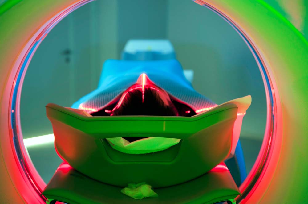
During the Scan
- You’ll lie on a narrow table that slides into the scanner
- The technologist will leave the room but can see and hear you throughout
- You’ll need to lie still while the machine takes images
- The table will move several times during the scan
- You’ll hear buzzing sounds as the X-ray tube rotates around you — these sounds aren’t as loud as an MRI but may feel unsettling if it’s your first time
- The entire process usually takes 10 to 30 minutes
After the Scan
- You can typically resume normal activities immediately
- Drink plenty of water to help flush the contrast material from your body
- Your doctor will discuss the results with you after a radiologist interprets the images
Can a CT Scan Show Ovarian Cancer Spread?
One of the greatest strengths of CT scans is their ability to show if ovarian cancer has spread beyond the ovaries. This crucial information directly determines your treatment options.
CT scans effectively show:
- Tumor spread to nearby pelvic organs
- Cancer in the lymph nodes
- Metastases to the liver, lungs, or other distant organs
- Fluid buildup in the abdomen (ascites), which commonly occurs with ovarian cancer
The research indicates that CT scans are particularly accurate in detecting disease in certain areas, such as the diaphragm and omentum (fatty tissue covering the abdominal organs), but less reliable in showing the involvement of gastrointestinal organs and the mesentery, a membrane that attaches the intestine to the stomach wall.
Comparing CT Scans to Other Diagnostic Methods
Ovarian cancer CT scans work alongside several other diagnostic tools. Here’s a quick comparison:
Transvaginal Ultrasound: Usually the first test ordered; better for examining ovarian structure; no radiation; limited in showing cancer spread.
MRI: Provides more detailed soft tissue images than CT; better at determining if masses are benign or malignant; no radiation but more expensive and less available.
PET/CT Scan: Highlights metabolically active areas like cancer; excellent for detecting recurrence but less useful for initial diagnosis.
CA-125 Blood Test: Measures a protein often elevated with ovarian cancer; used alongside imaging for diagnosis and monitoring treatment response. Learn more about CA125 ovarian cancer screening.
When ultrasound results are unclear, MRI typically provides the most reliable assessment of whether an ovarian mass might be cancerous.
When Is a Surgical Evaluation Needed?
While imaging tests like CT scans provide valuable information, they cannot definitively diagnose ovarian cancer. Only a biopsy — removing tissue and examining it under a microscope — can confirm cancer.
If your CT scan and other tests suggest possible ovarian cancer, your doctor will likely refer you to a gynecologic oncologist — a specialist in cancers of the female reproductive system. This specialist may recommend:
- A laparoscopy (minimally invasive surgery) to visually examine the ovaries and take biopsies
- A laparotomy (open surgery) for more extensive evaluation and possible tumor removal
Research shows that surgeries performed by gynecologic oncologists can result in survival rates as much as 30% greater compared to those performed by less experienced surgeons, including general surgeons, benign gynecologists, or general OB/GYNs.
Using CT Scans to Guide Treatment
Beyond diagnosis, CT scans play a vital role in treatment planning:
Surgical Planning: CT helps surgeons understand the extent of disease before operating. The scan shows which organs are affected and whether complete surgical removal of the cancer is achievable.
Treatment Selection: CT findings help determine whether a patient should undergo immediate surgery or receive chemotherapy first.
Treatment Monitoring: During and after treatment, repeat CT scans evaluate how well the treatment is working.
Research indicates that CT scans are the preferred technique for pretreatment evaluation of ovarian cancer to define disease extent and assess the likelihood of optimal surgical cytoreduction (removal of as much tumor as possible).
Moving Forward After Your CT Scan
If your CT scan indicates possible ovarian cancer:
- Request a referral to a gynecologic oncologist
- Bring a support person to appointments to help process information
- Prepare specific questions about next steps and treatment options
- Obtain copies of your imaging reports and other test results
- Consider seeking a second opinion, especially for complex cases
Final Thoughts
Can a CT scan detect ovarian cancer? Yes, but it works most effectively as part of a comprehensive diagnostic approach. CT scans excel at showing larger tumors and cancer spread, making them invaluable for treatment planning. However, they have real limitations in detecting early-stage disease and determining whether masses are cancerous or benign.
If you’re experiencing persistent symptoms that concern you, don’t wait. Talk to your doctor promptly about your symptoms and the appropriate next steps. While ovarian cancer diagnosis presents challenges, advances in imaging technology continue to enhance our ability to detect and treat this disease effectively.