
Transvaginal Ultrasound: What It Shows, What It Can't, What to Expect
September 3, 2026

A transvaginal ultrasound is sometimes the first imaging test doctors order when something looks off in the pelvis, and for many women it is the moment the words “ovarian cancer” first enter the conversation.
This guide covers what the procedure is, what it can and can’t tell you, and what actually happens in that room.
Sign up to receive vital updates through email, and learn how you can get involved. "*" indicates required fieldsStay informed
What Is a Transvaginal Ultrasound?
A transvaginal ultrasound is an imaging test that uses high-frequency sound waves to create pictures of your uterus, cervix, endometrium, fallopian tubes, ovaries, and bladder.
A thin instrument called a transducer goes a few inches into the vagina, sends out sound waves, and captures the echoes that bounce back off your tissues. A computer converts those echoes into a moving image on a screen. No radiation. No needles. No incisions.
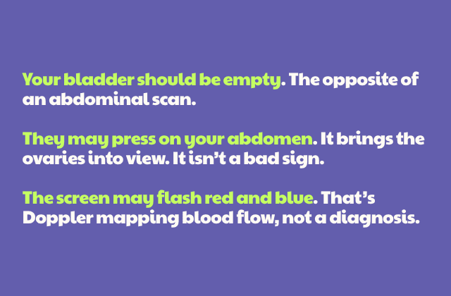
Two things about this test surprise people, so it’s worth saying up front. First, you’ll usually be asked to empty your bladder right before the scan. That’s the opposite of a transabdominal ultrasound, where a full bladder acts as a window into the pelvis. Here, the probe sits close to the organs already, and an empty bladder gets it closer still, giving a sharper view of the ovaries and uterus.
Second, the sonographer may press down on your lower abdomen with their free hand during the scan. Ovaries move, and gentle external pressure nudges them into the probe’s field of view. Expect it, and it feels routine instead of alarming.
You’ll also see this test called transvaginal sonography, endovaginal ultrasound, or simply TVUS.
The Transvaginal Ultrasound Probe
 People worry about the probe more than anything else, so let’s be specific.
The transvaginal ultrasound probe is a smooth, rounded wand designed for the curve of the vagina. It’s typically less than 1.5 inches in diameter (can range between 0.7 to 0.9 inches), which is wider than a tampon but noticeably smaller than the speculum used during a Pap test.
Before insertion, the sonographer covers it with a sterile protective sheath and coats it with lubricating gel. Only the tip of the probe goes in. It gets angled and rotated gently during the scan to capture different planes.
What Does a Transvaginal Ultrasound Show?
Quite a lot, and this is where it earns its place in an ovarian cancer workup. A transvaginal ultrasound can show:
- The size, shape, and position of your ovaries and uterus
- Ovarian cysts, including whether they’re simple fluid-filled sacs or something more complex
- Solid masses and papillary projections growing into a cyst
- Fibroids, polyps, and endometrial thickening
- Blood flow within a mass, measured with Doppler
- Free fluid in the abdomen, which can signal ascites
- Whether an IUD sits where it should
That Doppler piece deserves a note, because it happens during the same appointment and most patients never hear it explained. Color Doppler maps the movement of blood through tissue, and the sonographer may switch it on partway through the scan.
You might see patches of red and blue appear on the screen, or hear a whooshing sound if they turn on the audio. The colors indicate direction of flow. Tumors tend to build their own blood supply, so a mass with vessels running through it, particularly through its solid parts, reads differently than one with none.
What the scan shows about a mass matters enormously. Radiologists look at volume and morphology together. A cyst with smooth walls and clear fluid behaves very differently from a mass with solid areas, thick septations, and blood flow running through it.
But here is the honest limit, and every source worth trusting says the same thing: an ultrasound cannot tell you whether a mass is cancer. It can flag risk. It cannot confirm a diagnosis. Only tissue, examined by a pathologist, does that.
Why Your Doctor Ordered One
Most transvaginal ultrasounds are ordered because of symptoms: persistent bloating, pelvic pain in women that won’t quit, abnormal or postmenopausal bleeding, feeling full after a few bites, and needing to urinate constantly.
Those are also the ovarian cancer symptoms most often dismissed as digestive trouble or stress, which is exactly why the test matters. It can also be used to follow a known cyst over time, to check the ovaries before surgery, and to monitor patients already in treatment.
Preparing for a Transvaginal Ultrasound
Preparation for a transvaginal ultrasound is minimal, and most of it takes about five minutes of planning.
How to prepare:
- Wear something you can slip out of easily from the waist down. You’ll likely change into a gown.
- Remove a tampon before the exam if you’re menstruating. (You can still have the test during your period, and for some gynecologic questions that timing gives clearer information.)
- Bring your physician’s order and your insurance card if your center requires them.
- Leave jewelry and valuables at home.
If you’re anxious about the exam, say so when you check in. Staff can walk you through each step before anything begins, and knowing what’s coming makes a real difference.
What Happens During the Exam
 You’ll change into a gown and lie on your back on the exam table, knees bent, feet possibly in stirrups, the same position as a pelvic exam. A sheet covers you.
The sonographer inserts the covered, lubricated probe and moves it gently to capture images from different angles. You may feel pressure. They may press on your lower abdomen with a free hand to bring your ovaries closer to the probe.
Sometimes the radiologist steps in to review images or rescan an area. That’s routine, not a red flag.
You can ask for a chaperone. You can ask for a female sonographer, though availability varies and may require rescheduling. You can ask them to stop at any point. These aren’t special favors. They’re standard practice.
Making Sense of Your Results
Many radiologists now report adnexal findings using O-RADS, the Ovarian-Adnexal Reporting and Data System developed by the American College of Radiology. It assigns a score from 1 to 5 based on the mass’s appearance, with each category tied to an estimated risk of malignancy and a suggested next step. (0 means they couldn’t assess the mass.)
Low scores often mean watchful waiting with a repeat scan. Higher scores prompt referral, further imaging, or surgical evaluation.
A CA-125 blood test frequently accompanies the ultrasound. CA-125 rises in endometriosis, fibroids, pelvic inflammatory disease, liver disease, and even during menstruation, and plenty of early-stage cancers never elevate it at all.
If your results raise concern, ask for a referral to a gynecologic oncologist rather than proceeding with a general surgeon or generalist OB-GYN. Specialist care is associated with better outcomes. If your results come back normal but your symptoms persist, inquire further.
Why Transvaginal Ultrasound Isn’t a Routine Screening Test
A lot of women finish a transvaginal ultrasound and ask a reasonable follow-up question: if this test sees my ovaries so clearly, why isn’t it part of my annual checkup?
It’s been tried, at enormous scale, and it didn’t save lives. One study followed more than 200,000 women with no symptoms and no family history for an average of 16 years, comparing annual screening against no screening at all.
The final results found that screening caught some cancers earlier but didn’t reduce deaths from the disease. It also flagged plenty of findings that turned out to be nothing, sending some women into surgery to remove healthy ovaries. U.S. preventive health guidelines now advise against routine ovarian cancer screening for women without symptoms or known genetic risk.
None of that applies once you have symptoms. Ultrasound and CA-125 remain proven parts of the diagnostic workup. The question was never whether these tools work, only whether they work as a net cast over healthy women.
And timing is everything here. Caught while it’s still confined to the ovary, invasive epithelial ovarian cancer has a five-year relative survival rate of 92%. Once it has spread, that drops to 32%. Which is why persistent symptoms deserve a real workup instead of wait-and-see.
Frequently Asked Questions
Let’s answer some common questions about having a transvaginal ultrasound.
Is a Transvaginal Ultrasound Painful?
For most people, no. Uncomfortable, sometimes. Painful, usually not.
The probe is shaped and lubricated to slide in without resistance, and the pressure sensation typically fades within seconds. Some patients experience light spotting or mild cramping afterward, which resolves quickly.
That said, discomfort isn’t evenly distributed, and this test isn’t for everyone. It’s generally avoided in patients who aren’t sexually active, and it should never proceed over an objection. If you decline vaginal insertion for any reason, that’s a complete answer, and no explanation is owed.
Recent vaginal surgery may rule the test out entirely. In those situations, providers can offer alternatives such as a transabdominal ultrasound, CT, or MRI. Our guide on can a CT scan detect ovarian cancer covers what those other options do and don’t catch.
Tell your provider beforehand if you have concerns.
How Long Does a Transvaginal Ultrasound Take?
The scan itself generally runs 15 to 30 minutes, depending on what’s being examined and how easily your anatomy comes into view. Some centers quote 15 to 20 minutes for a straightforward exam, while others build in 30 to 60 minutes for the full appointment including setup and any additional images.
Plan for about an hour at the facility. You can return to normal activity immediately afterward.
Results take longer than the scan. A radiologist reviews the images and sends a report to the ordering physician, typically within a few hours to a few days. The waiting is often the hardest part, and if that stretch of time is eating you alive, our piece on scanxiety offers practical strategies.
Pelvic Ultrasound vs. Transvaginal Ultrasound
“Pelvic ultrasound” is the umbrella term. It refers to any ultrasound imaging of the female pelvic organs, and it’s performed one of two ways:
- Transabdominal: gel on the lower abdomen, probe moved across the skin, usually requiring a full bladder. Wider field of view, lower resolution.
- Transvaginal: probe inserted into the vagina. Narrower field of view, much higher resolution.
So a transvaginal ultrasound is a pelvic ultrasound.
Oftentimes, both get done in one appointment, and there’s a good reason for that. The transvaginal probe gives a detailed view of the ovaries but covers a narrow field. A large mass sitting higher in the abdomen could fall outside its view entirely. The abdominal scan catches what the internal scan might miss. Between the two, you get detail and coverage. For a broader look at how imaging fits the whole diagnostic picture, see our ovarian cancer ultrasound guide.
Can a Transvaginal Ultrasound Detect Ovarian Cancer?
It can detect abnormalities that suggest cancer, including complex masses, solid components, and free fluid. It cannot confirm cancer. Diagnosis requires tissue evaluation, usually after surgery.
Is a Transvaginal Ultrasound Safe?
Yes. It uses sound waves, not radiation, and has been used safely for decades
Can I Have the Test During My Period?
Yes. Remove your tampon first. For certain gynecologic questions, scanning during menstruation actually gives clearer information.
What if the Sonographer Finds Something?
Repeat scans are common, and the interval varies depending on the size and appearance of what was found.
Many cysts resolve on their own or stay stable, so follow-up can be scheduled anywhere from a few weeks to a year out. Under O-RADS, a simple cyst classified as almost certainly benign may not need rechecking for months, while larger or less clear-cut findings get a shorter leash.
Persistent or complex findings usually trigger a CA-125 test, additional imaging, and a specialist referral.
Does a Transvaginal Ultrasound Show the Fallopian Tubes?
It images the tubes, but standard scanning is not reliable for detecting the small precancerous changes now understood to precede many serous ovarian cancers.
Will it Hurt if I’ve Never had Penetrative Sex?
It may be more uncomfortable, and it isn’t always appropriate. Tell your provider, and you may ask about transabdominal imaging or MRI instead.
Do I Need Someone with Me?
Not medically. But you’re allowed to bring a family member or friend, and you can request a chaperone regardless of the sonographer’s gender.
One Test, Not the Whole Answer
A transvaginal ultrasound is a fast, safe, unusually informative test, and it’s a reasonable place for your workup to start. It is also, on its own, an answer that stops short of being an answer.
If you’re waiting on results right now, write down your questions before your follow-up appointment. Our list of ovarian cancer questions to ask your doctor will help you make that conversation count.